Building a Preclinical Procedure Station
What labs need for anesthesia, oxygen, monitoring, and surgery
TL;DR
A good preclinical procedure station is built around the procedure, species, endpoint, and user workflow. Start with anesthesia delivery and oxygen strategy, then plan respiratory support, monitoring, surgical tools, warming, recovery, documentation, and training. The goal is a station that protects the animal, supports cleaner data, and helps every user run the workflow the same way.
Build your station around the study
Most labs do not buy equipment because they want more equipment.
They are trying to run a procedure consistently.
That is the real job of a preclinical procedure station. It should help the team move from induction to procedure to recovery with fewer variables, fewer surprises, and fewer places where the workflow depends on memory or improvisation.
The station may include an anesthesia system, oxygen source, intubation tools, endotracheal tubes, nebulizer, ventilator, pulse oximeter, blood pressure system, warming support, forceps, hemostats, surgical platform, and recovery setup.
That sounds like a lot.
The cleaner way to think about it is sequence:
What does the animal need before, during, and after the procedure?
Start there. Then choose the tools.
Start with species, endpoint, and procedure length
A mouse imaging study, rat survival surgery, stereotaxic procedure, cardiovascular workflow, and respiratory model do not need the same station.
Before choosing equipment, define:
- species and strain
- body size
- procedure length
- anesthetic agent
- oxygen strategy
- airway needs
- monitoring requirements
- warming plan
- sampling needs
- recovery requirements
- who will run the workflow
This planning step protects the whole study.
The National Academies’ Guide for the Care and Use of Laboratory Animals notes that monitoring during anesthesia includes anesthetic depth and physiologic functions such as body temperature, cardiac and respiratory rates and pattern, and blood pressure, with appropriate documentation. See the National Academies guidance here: Guide for the Care and Use of Laboratory Animals.
It’s a useful frame for building your station. Your equipment should support what your animal and procedure require.
Anesthesia delivery sets the workflow
Anesthesia is often treated like setup.
In practice, anesthesia shapes the whole procedure: positioning, access, warming, respiratory support, monitoring, user workflow, waste gas control, and recovery.
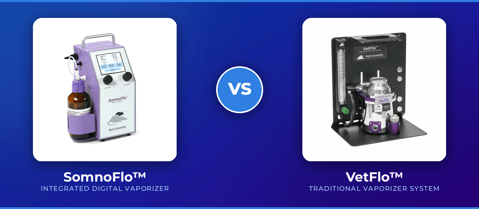
A traditional vaporizer may work well in a lab with stable users, clear SOPs, appropriate service, and strong flow discipline. Integrated low-flow systems may be a better fit when the lab needs small animal-specific delivery, lower gas use, and more repeatable settings across users.
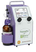
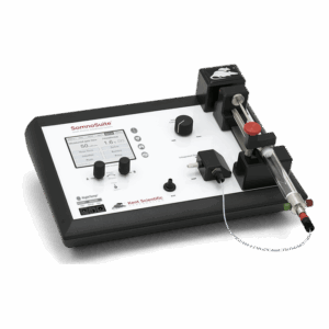
Kent’s SomnoSuite® is a low-flow anesthesia system designed for mice and rats. Kent’s SomnoFlo® and SomnoFlo® O2Care support low-flow electronic vaporization for small animal anesthesia workflows.
When choosing an anesthesia system, ask:
- How long is the animal under anesthesia?
- How many users run the station?
- Does the lab need one animal or multiple animals under anesthesia?
- Does the procedure require oxygen blending?
- Is the workflow tied to imaging, stereotaxic surgery, ventilation, or physiologic monitoring?
- How easy is it to train a new user?
Those questions matter more than the equipment category alone.
Oxygen source: tank, concentrator, or blended oxygen
Oxygen strategy belongs early in the station design and depends on both study aims and institutional policies.
Some protocols use compressed oxygen tanks. Some use an oxygen concentrator. Some use room air alone, and some add supplemental oxygen. Some systems allow oxygen blending so the team can select a defined oxygen concentration for the protocol.
A tank can be straightforward and familiar, but it requires supply management, regulator checks, and storage planning.
An oxygen concentrator can reduce dependence on cylinders, but the lab still needs to confirm flow capacity, output concentration, compatibility with the anesthesia setup, and facility requirements.
A blended oxygen system gives the team more control over oxygen concentration when the procedure or model requires it. Typically, an external gas mixer is used for convenience and consistency, but these come with similar drawbacks to tanks and concentrators: supply management, storage planning, facility requirements, and compatibility with anesthesia setup.
The internal gas mixer in the SomnoFlo O2Care is designed for low-flow anesthesia to deliver adjustable oxygen concentrations for small animal workflows through Kent’s SomnoFlo O2Care product page.
The practical question:
What oxygen strategy supports the animal, endpoint, and protocol without adding unnecessary complexity?
Write the answer into the SOP.
Respiratory support starts with tidal volume
Tidal volume is the amount of air moved in or out of the lungs with each breath.
In small animals, the scale is small. That makes respiratory support sensitive to equipment choice and user technique.
A reference table from Biology of the Laboratory Mouse lists mouse tidal volume at about 0.15 mL, with a reported range of 0.09–0.23 mL depending on sex, strain, and weight. See the Jackson Laboratory table here: Respiratory Characteristics of Mice.
That small volume is one reason rodent anesthesia and ventilation workflows need size-appropriate equipment.
Respiratory planning may include:
- anesthetic type and how it affects spontaneous breathing
- assisted ventilation, including factors associated with intubation
- specific respiratory rate and tidal volume needs
- oxygen strategy (even with injectable anesthetics!)
- waste gas control
- recovery monitoring
If the procedure may affect breathing, or if the animal will be under anesthesia for a longer window, respiratory support should be planned before the first cohort begins.
Ventilation, intubation and endotracheal tubes: a right size plan before the animal is on the table
Ventilation is not only for large animals.
Rodent workflows may require ventilation when spontaneous breathing is insufficient, when the procedure affects respiratory function, or when controlled ventilation supports the study design. In small animal research, intubation and ventilation can be helpful for workflows that need controlled parameters, longer anesthesia duration, access to the head or face, or more direct respiratory support. It can also add training requirements and technical variability.
Kent’s RoVent® and RoVent® Jr. systems are designed for small animal ventilation workflows. Product choice depends on species, animal size, procedure type, and the level of control
The station should support the airway plan. Ventilation planning should include:
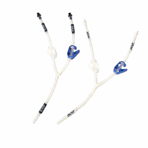
- Appropriately sized endotracheal tubes for species and weight range
- tidal volume target
- respiratory rate
- inspiratory and expiratory timing
- Anesthetic choice (inhalant or injectable)
- oxygen source or other carrier gas (if needed)
- Airway access method, including visualization tools and positioning aids when necessary
- Monitoring, including tube placement and warming support
- recovery plan
- Documentation of tube size and ventilation settings
If the lab will intubate mice or rats, training becomes part of the equipment plan. If the station includes a ventilator, the SOP should explain when to use it, who is trained to use it, what settings are allowed, and how the animal is monitored. Small animal ventilation should be treated as a workflow, not an accessory.
A ventilation setup that only one expert can run well is fragile. A station that supports clear positioning, visibility, and repeatable steps is easier to teach and easier to defend.
Where a nebulizer fits in the station
A nebulizer creates an aerosol from liquid, often for respiratory exposure or delivery workflows.
In preclinical research, nebulizers may be used in respiratory models, inhalation studies, compound delivery, airway irritation studies, infectious disease workflows, or other protocol-specific applications.
Nebulizer use changes the station.
It may affect:
- containment
- exposure control
- animal restraint or housing
- respiratory monitoring
- cleaning
- dose consistency
- staff safety
- documentation
A nebulizer should be planned as part of the respiratory workflow, not added at the end.
Ask:
- Is the nebulizer used for delivery, exposure, humidification, or another protocol-specific purpose?
- Does the workflow require anesthesia?
- Does the animal breathe spontaneously or require airway support?
- How is aerosol contained?
- How is the station cleaned between animals or groups?
- How is dose or exposure time documented?
Those questions help keep the nebulizer from becoming an uncontrolled variable.
Monitoring turns the station into a feedback system
Monitoring helps the team see when the procedure is drifting.
The Guide for the Care and Use of Laboratory Animals notes that monitoring can include anesthetic depth, body temperature, cardiac and respiratory rates and pattern, and blood pressure, with appropriate documentation. See the monitoring discussion here: Guide for the Care and Use of Laboratory Animals.
That is the purpose of an intraopreative monitoring workflow.
It should help users answer:
- Is the animal stable?
- Is the procedure affecting physiology?
- Is warming working?
- Is breathing changing?
- Is blood pressure part of the endpoint?
- Is recovery on track?
Monitoring does not need to be complicated. It needs to match the procedure.
Pulse oximetry: useful signal, careful interpretation
A pulse oximeter can help monitor oxygen saturation and pulse-related signals in small animal workflows.
In rodent research, pulse oximetry can support anesthesia monitoring, oxygenation checks, procedure stability, and recovery observation. It can also be affected by sensor placement, animal movement, perfusion, temperature, pigmentation, and signal quality.
Kent’s MouseSTAT® Jr. is designed for pulse oximetry and heart rate monitoring in mice and rats as part of small animal physiologic monitoring workflows.
A pulse oximeter should be used as part of a broader monitoring plan.
Ask:
- Where will the sensor be placed?
- Will the animal be warmed?
- Is perfusion adequate?
- Will the animal move?
- How will poor signal quality be handled?
- What gets recorded?
The goal is not to collect a number. The goal is to collect a number the team can interpret.
Temperature belongs in every anesthesia plan
Small animals can lose heat quickly under anesthesia.
Temperature drift can affect recovery, physiology, and data quality. It can also make other monitoring signals less reliable.
A warming plan should cover the full workflow:
- induction
- positioning
- procedure
- transfer
- recovery
Kent’s FIR Warming systems, including the RightTemp® and RightTemp® Jr., as well as the SurgiSuite are designed to help support thermal control in small animal workflows.
Temperature should not be added only when the animal looks cold.
It should be part of the station design.
Blood pressure monitoring: where CODA fits
Blood pressure can be an endpoint, a safety measure, or a physiologic variable that affects interpretation.
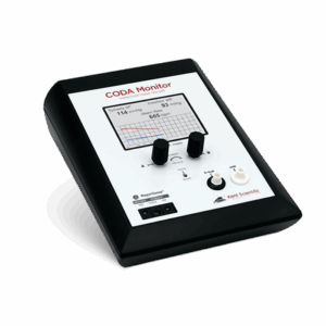
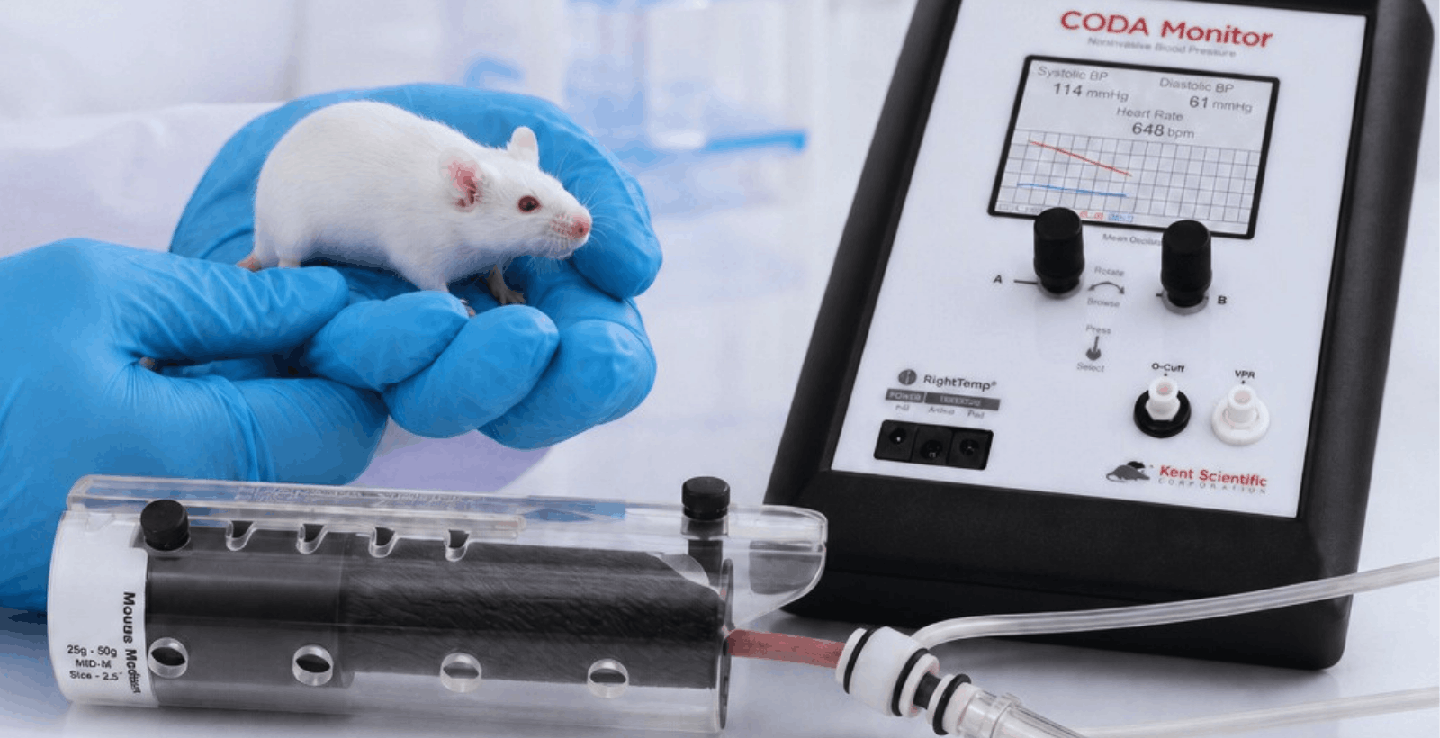
Kent’s CODA® non-invasive blood pressure systems use tail-cuff technology for blood pressure measurement in mice and rats. CODA can support cardiovascular, toxicology, pharmacology, hypertension, renal, metabolic, and related workflows where non-invasive blood pressure data matters. The CODA can expand beyond the surgical suite and can also be used to collect data in awake animals; perfect for pre- or post-operative monitoring… or validating your hypothesis!
For tail-cuff blood pressure, consider:
- Anesthetic type
- warming requirements
- cuff size
- training
- frequency measurements
- data recording
- user consistency
A CODA station should be built around repeatability. At the end of the day, tail-cuff technology only works if there’s enough blood in the animal’s tail, which can be affected by stress, temperature, movement, handling, anesthetic type, and procedure length; no matter if you take a reading pre-, peri-, or post-operatively, these should factor into your workflow.
Surgical tools: forceps and hemostats are part of the workflow
In rodent surgery, instrument choice affects tissue handling, procedure time, access, and recovery. Forceps and hemostats look simple until they are the wrong size, wrong pattern, or wrong fit for the tissue and procedure.
Forceps may be used for delicate handling, tissue positioning, support, or microdissection. Hemostats may be used for clamping, holding, or controlling bleeding where appropriate.
The right surgical setup depends on:
- species
- tissue type
- procedure depth
- incision size
- access angle
- surgeon training
- survival versus terminal procedure
- need for atraumatic handling
- cleaning and sterilization workflow
A rodent surgery station should make the procedure easier to run carefully.
That means instruments, platform, warming, lighting, magnification, positioning, anesthesia access, and recovery support all need to work together.
Kent’s SurgiSuite is designed as a small animal surgical platform with warming, lighting, magnification, and positioning support for rodent workflows.
Positioning and warming shape the surgical field
A stable surgical field helps the animal and the user.
Positioning affects access, airway alignment, anesthesia delivery, warming contact, monitoring sensor placement, and procedure time.
For rodent procedures, the station should support:
- stable body position
- appropriate restraint
- access to the surgical site
- nose cone or airway access
- warming surface contact
- clean fluid management
- visibility
- repeatable setup between users
Small positioning differences can create large workflow differences.
If one user tapes differently, another places the nose cone differently, and another changes warming position, the procedure may become harder to reproduce.
A good station reduces those small variations.
Recovery is part of the procedure station
Recovery is often where weak setup becomes visible.
An animal that recovers slowly, cools down, breathes unevenly, or needs more intervention may be showing the downstream effects of anesthesia depth, temperature loss, procedure time, respiratory support, or handling.
Recovery planning should include:
- warming
- observation
- oxygen support if required by protocol
- documentation
- return-to-cage criteria
- monitoring frequency
- who is responsible for post-procedure checks
Institutional guidance often emphasizes monitoring animals through recovery after anesthesia. For example, the University of Iowa’s anesthesia guidance lists recovery monitoring considerations such as body temperature, heart and respiratory rate, and observation until appropriate recovery milestones are reached. See: University of Iowa IACUC anesthesia guidance.
Your institution may use different criteria. The important point is consistent planning.
Recovery should be included in procedure planning… long before the animal is induced.
Documentation keeps the station defensible
A well-designed station still needs records.
Documentation helps the lab show what happened, who ran the procedure, what settings were used, what was monitored, and whether the animal recovered as expected.
Record:
- animal ID
- species and weight
- anesthetic agent
- induction settings
- maintenance settings
- oxygen source and concentration
- ventilation settings, if used
- temperature support
- pulse oximetry or other applicable monitoring data
- blood pressure data, if relevant
- surgical tools or implants, if relevant
- complications
- recovery observations
The UCSF IACUC notes that complete surgery and anesthesia recordkeeping helps demonstrate protocol compliance and animal welfare, while also helping detect trends in surgical or anesthetic outcomes. See: UCSF surgery and anesthesia recordkeeping for rodents.
Good documentation is not busywork.
It is how the station becomes repeatable, and your data becomes more reliable.
What to standardize before the first cohort
A preclinical procedure station should be standardized before the study begins.
Do not wait for the first animal to reveal the gaps.
Standardize:
- anesthesia setup
- oxygen source
- induction and maintenance settings
- airway plan
- ventilation criteria
- monitoring plan
- warming setup
- surgical tools
- positioning method
- recovery setup
- documentation fields
- cleaning and turnover
- user training
- service and certification schedule
Then run a pilot or dry setup where appropriate.
That is how a lab finds missing connectors, wrong cuff sizes, incompatible tubing, awkward positioning, unclear SOP language, or training gaps before they affect the study.
When to call Kent before building the station
Call Kent when the workflow involves more than one moving part.
That includes:
Kent can help map the station from induction through recovery so the equipment supports the study instead of becoming another variable.
Example station maps
Mouse survival surgery station
A typical mouse survival surgery station is built for the procedure, and may include low-flow anesthesia, oxygen source, nose cone or ventilation equipment plan, warming support, surgical platform, forceps, hemostats, pulse oximetry where appropriate, recovery warming, and documentation.
Kent fit: SomnoSuite or SomnoFlo/o2 care, RightTemp, SurgiSuite, MouseSTAT Jr., surgical tools, PhysioSuite, RoVent, and Kent SC service.
Rat cardiovascular monitoring station
If you’re just monitoring cardiovascular parameters without surgery, a rat cardiovascular station may include anesthesia or awake measurement planning, warming, CODA blood pressure monitoring, oxygen strategy, restraint or acclimation plan, and consistent documentation.
Kent fit: CODA, warming tools, MouseSTAT Jr., PhysioSuite, SomnoSuite or SomnoFlo
Respiratory support station
A respiratory support station may include anesthesia, oxygen source or concentrator, endotracheal tube, intubation tools, ventilator, appropriate monitoring, warming, and recovery monitoring.
Kent fit: SomnoFlo O2Care, SomnoSuite, RoVent or RoVent Jr., MouseSTAT Jr., RightTemp, Physiosuite (especially CapnoScan module), mouse/rat intubation kit, and other accessories
Surgical teaching station
A teaching station should reduce user-to-user variability, independent of what procedures you’re teaching. It may include a standardized anesthesia setup, fixed positioning method, warming, a defined instrument kit, forceps, hemostats, monitoring guidance, recovery checklist, and required record fields.
Kent fit: SurgiSuite, surgical tools, SomnoSuite or SomnoFlo, RightTemp, MouseSTAT Jr., PhysioSuite.
Build once. Run consistently.
A preclinical procedure station should make the right workflow easier to repeat.
That is the real value.
The best station is not the one with the most equipment. It is the one that supports the animal, the endpoint, the user, and the record.
Start with the procedure.
Map the workflow.
Choose the tools.
Train the users.
Document the setup.
Then run it the same way every time.
That is how a station protects the animal, the data, and the study timeline.
Talk to Kent about your procedure station
If you are building or updating a preclinical procedure station, Kent can help you think through the workflow before you buy the wrong mix of equipment.
We can help with anesthesia, oxygen strategy, monitoring, ventilation, blood pressure, warming, surgical setup, service, and training.
Talk to Kent about your workflow
Explore anesthesia systems
Explore monitoring systems
Schedule service or training
FAQ: Building a preclinical procedure station
What is a preclinical procedure station?
A preclinical procedure station is the equipment and workflow setup used to run animal procedures consistently. It may include anesthesia, oxygen delivery, ventilation, monitoring, warming, surgical tools, positioning, recovery support, and documentation.
What equipment does a rodent anesthesia station need?
It depends on the procedure! A rodent anesthesia station may include an anesthesia delivery system, oxygen source, induction chamber, nose cone or intubation setup, scavenging, warming support, monitoring, and recovery area. The exact setup depends on species, procedure length, endpoint, and institutional protocol.
When does a lab need an endotracheal tube or intubation setup?
A lab may consider intubation and an endotracheal tube when the procedure requires airway access, controlled ventilation, longer anesthesia, or respiratory support. The workflow should be planned with training, monitoring, and institutional veterinary guidance.
Why does tidal volume matter in small animal ventilation?
Tidal volume is the volume of air moved with each breath. Small animals have very small tidal volumes, so ventilation equipment and settings need to be scaled appropriately. A mouse reference table from Biology of the Laboratory Mouselists tidal volume around 0.15 mL. Respiratory Characteristics of Mice
What does a pulse oximeter measure in small animal research?
A pulse oximeter can help monitor oxygen saturation and pulse-related signals. In small animals, signal quality can be affected by sensor placement, temperature, movement, perfusion, and animal size.
Where does CODA fit in a preclinical station?
CODA fits in workflows that need non-invasive blood pressure monitoring in mice or rats. It is often used in cardiovascular, toxicology, pharmacology, hypertension, renal, metabolic, and related studies.
What surgical tools should be included in a rodent surgery station?
Common tools include forceps, hemostats, scissors, needle holders, retractors, clamps, sutures, and procedure-specific instruments. Tool selection should match species, tissue type, procedure depth, survival status, and user training.
Does a nebulizer belong in a preclinical procedure station?
A nebulizer may belong in the station when the protocol requires aerosol delivery, respiratory exposure, inhalation workflows, or airway-related studies. Nebulizer use should be planned around containment, dose consistency, cleaning, respiratory monitoring, and staff safety.
What procedure station settings should be documented?
Labs should document species, weight, anesthetic agent, induction and maintenance settings, oxygen strategy, airway method, ventilation settings, monitoring data, warming setup, surgical notes, complications, and recovery observations.
Check out this blog for tips on limiting anesthesia gas waste