Veterinary handheld ultrasound scanner for skin assessment, monitoring, and documentation
SkinScanner combines high-frequency ultrasound and dermoscopy-assisted imaging to help veterinary teams evaluate skin lesions, monitor changes over time, and support more consistent clinical decisions.
Built for veterinary hospitals, specialty clinics, and dermatology-focused care
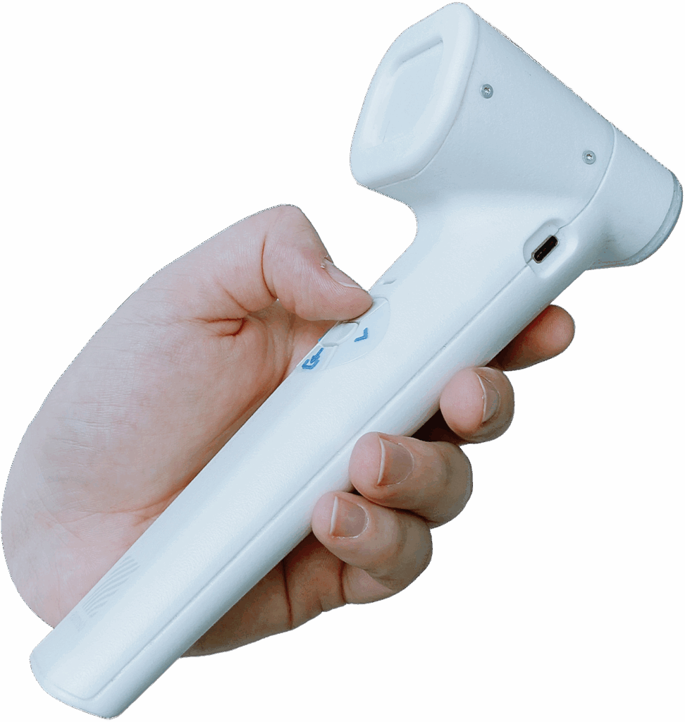
A veterinary handheld ultrasound scanner designed for skin and superficial tissue imaging, with reproducible positioning and structured documentation built into the workflow.

20–40 MHz handheld ultrasound

Dermoscopy-assisted positioning

Up to 10 mm imaging depth
See deeper in veterinary skin cases
When a patient presents with a visible lesion, mass, or area of irritation, surface inspection only tells part of the story. Veterinary teams need a better way to examine skin and superficial tissue, document what they see, and compare findings across rechecks.
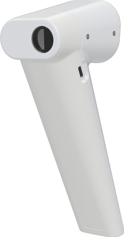
SkinScanner is the veterinary handheld ultrasound scanner built for practices that want more than photos and chart notes.
It helps clinicians capture reproducible images, assess structures below the surface, and create a stronger clinical record over time.
Why do veterinarians use SkinScanner handheld ultrasound scanner for skin imaging?
Skin cases get complicated when the record is limited to visual inspection.
The SkinScanner handheld ultrasound scanner for veterinarians gives your team a more structured way to evaluate skin lesions and superficial abnormalities.
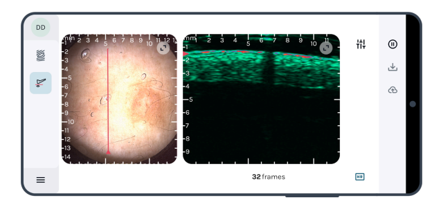
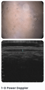
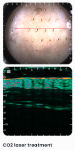
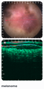
With high-frequency imaging and dermoscopy-guided positioning, SkinScanner helps Dermoscopy-assisted multimodal imaging guidance with a 12 × 12 mm optical field of view.
That means better monitoring, clearer documentation, and fewer shrug emoji moments in the exam room.
See more with the highest frequency handheld ultrasound
SkinScanner
A high-frequency veterinary handheld ultrasound scanner designed for evidence-based skin evaluation and treatment monitoring.
- Highest frequency handheld ultrasound on the market: 20–40 MHz
- Imaging depth up to 10 mm
- Dermoscopy-assisted guidance with a 12 × 12 mm optical field of view
- High-resolution and high-depth imaging modes
- Easy interpretation with positioning line
- Precise image positioning and reproducibility
- Power Doppler mode
- Affordable point-of-care format
SkinAid software
A web-based platform for image review, annotation, and skin change analysis.
- AI-based skin layer segmentation
- Quantification of skin changes
- Smart annotations, filters, and settings
- Third-party image import
- Two-point distance measurement and volume estimation
- Cloud or local storage options
- Efficient patient data management


Common skin scanning applications
Note: some features (such as automated segmentation and blood flow detection) are optimized for human applications.
SkinScanner supports veterinary teams evaluating and monitoring a range of skin-related findings, including:
- skin cancer:
- melanoma
- basal cell carcinoma
- squamous cell carcinoma
- skin inflammations:
- psoriasis
- atopic dermatitis
- hidradenitis suppurativa
- …and more
This makes it especially useful for practices managing dermatology, oncology, and repeat skin lesion follow-up.
Benefits for veterinary hospitals and specialty clinics
- Better repeatability
Dermoscopy-assisted positioning helps clinicians return to the same area for more consistent follow-up imaging.
- Better visibility below the surface
A veterinary handheld ultrasound scanner helps teams assess skin and superficial tissue beyond what is visible externally.
- Better owner communication
Share clearer findings with dermatology, oncology, surgery, or referral partners.
- Better referral support
Share clearer findings with dermatology, oncology, surgery, or referral partners.
- Better documentation
Organize images and measurements in one place for cleaner, more useful clinical records.
A better way to evaluate skin lesions
in veterinary care
SkinScanner gives your team a more precise way to assess skin findings, document changes, and support follow-up decisions with reproducible imaging.