Logo for Kent Scientific

Acrylic Brain Matrices

Acrylic Brain Matrices

Brain matrices were developed to provide medical researchers with the ability to precisely prepare discrete regions in fresh or preserved tissue samples.

Acrylic brain matrices are offered as durable and economical devices to prepare your tissue samples. Acrylic matrices are not recommended for heat or cold transfer and will become brittle if cooled or frozen.

We have selected our matrices to section tissue slices to a thickness as small as approximately 1 mm, depending upon what matrix design you choose to use. Ordinary industrial razor blades are generally used to slice the brain and/or tissue material. We recommend that the cutting blade be wetted with saline to act as a lubricant before shearing the target tissue. Micron tissue punches are also available for removal of specific areas of the tissues.

The coronal RBMs have the additional feature of a mid-line sagittal cut to facilitate the splitting of the left and right hemispheres. Sections can be as fine as 1 mm.

  • Repeatable slices to 1 mm providing precise blocking prior to sectioning
Your Price
Save for later

Cleaning Instructions

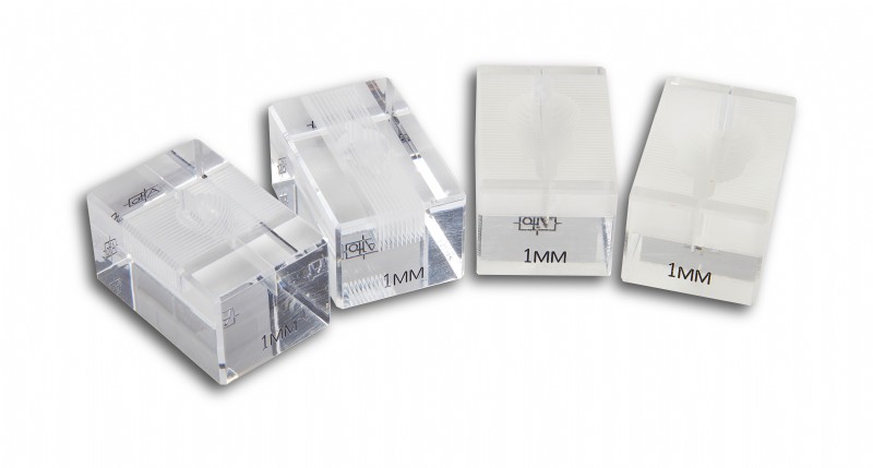
The acrylic material cannot be autoclaved and must be chemically sterilized.

Please note: Before using any chemical solutions, make sure the solution is compatible with the acrylic material. Place a drop or two of the solution on one side of your matrix and leave for a period of time to see if the sterilant causes any harm to the matrix. If the test is to your satisfaction, you can use the sterilant of choice. After using the sterilant to bathe the matrix, immediately rinse with clean water or saline solution. Do not immerse the matrix in the disinfectant, only wash and immediately rinse for best results.

Do not use any alcohol-based products on acrylic matrices.

Table 1: Recommended Hard Surface Disinfectants

Name Example Comments
Alcohols 70% ethyl alcohol
70% - 99% isopropyl alcohol
  • Contact time required is 15 minutes
  • Contaminated surfaces take longer to disinfect
  • Remove gross contamination before using
  • Inexpensive
  • Flammable
Quaternary Ammonium Roccal®, Cetylcide®
  • Rapidly inactivated by organic matter
  • Compounds may support growth of gram negative bacteria
Chlorine Sodium hypochlorite (Clorox ® 10% solution)
Chlorine dioxide (Clidox®, Alcide®)
  • Corrosive
  • Presence of organic matter reduces activity
  • Chlorine dioxide must be fresh (<14 days old)
  • Kills vegetative organisms within 3 minutes of contact
Aldehydes Glutaraldehyde (Cidex®, Cide Wipes®)
  • Rapidly disinfects surfaces
  • Toxic
  • Exposure limits have been set by OSHA
Phenolics Lysol®, TBQ®
  • Less affected by organic material than other disinfectants
Chlorhexidine Nolvasan®, Hibiclens®
  • Presence of blood does not interfere with activity
  • Rapidly bactericidal and persistent
  • Effective against many viruses

Table 2: Recommended Instrument Sterilants

Name Examples Comments
Physical: Steam Sterilization (Moist Heat) Autoclave Effectiveness dependent upon temperature, pressure and time (e.g., 121oC for 15 min. vs 131oC for 3 min)
Dry Heat Hot Bead Sterilizer, Dry Chamber Fast. instruments must be cooled before contacting tissue
Ionizing Radiation Gamma Radiation Requires special equipment
Chemical: Gas Sterilization Ethylene Oxide
  • Requires 30% or greater relative humidity for effectiveness against spores
  • Gas is irritating to tissue; all materials require safe airing time
  • Carcinogenic
Hydrogen Peroxide (Sterad®) Not useful for “delicate” items
Chlorine (inst. must be rinsed thoroughly with sterile water) Chlorine Dioxide (Clidox®, Alcide®)
  • A minimum of 6 hours required for sterilization
  • Presence of organic matter reduces activity
  • Must be freshly made (<14 days)
Aldehydes (inst. must be rinsed thoroughly with sterile water) Formaldehyde (6% sol.), Glutaraldehyde For all aldehydes:
  • Many hours required for sterilization
  • Corrosive and irritating
  • Consult safety representative on proper use
  • Glutaraldehyde is less irritating and less corrosive than formaldehyde

Matrix Dimensions - Adult Mouse 

  • A: 3.2 mm
  • B: 11.1 mm
  • C: 8.7 mm
  • D: 19.1 mm
  • E: 12.2 mm
  • Cavity Depth: 7.4 mm

Matrix Dimensions - Small Rat

  • A: 4.8 mm
  • B: 15.9 mm
  • C: 12.7 mm
  • D: 36.6 mm
  • E: 23.8 mm
  • Cavity Depth: 7.6 mm

Matrix Dimensions - Large Rat

  • A: 4.8 mm
  • B: 19.8 mm
  • C: 14.7 mm
  • D: 36.6 mm
  • E: 24.7 mm
  • Cavity Depth: 10.9 mm

Reference: The Mouse Brain in Stereotaxic Coordinates

BK-012547637X 
Deluxe 2nd Edition with CD ROM
Author: George Paxinos, Keith B.J. Franklin, Franklin Keith B. J.
Publication date: July 2001 / 296 pages / 2nd Edition

The Mouse Brain in Stereotaxic Coordinates is the most comprehensive and accurate atlas of the mouse brain ever published. The first edition of this book has become the acknowledged reference for the field. For this second edition, the authors have incorporated lower brainstem sections, added an entire sagittal plane section, and revised all delineations, especially of the cortex.

Key features:

  • A coronal set (slices are horizontal, from front to back) of 100 photographs and 100 matching detailed diagrams delineating the entire brain
  • A sagittal set (slices are vertical, from left to right) featuring 32 photographs and 32 matching detailed diagrams
  • Photographs of alternately Nissl- and Acetylcholinesterase-stained sections
  • Thoroughly revised cortical delineations and improved subcortical delineations
  • All diagrams available on CD-ROM

The CD-ROM contains:

  • The entire text of the Atlas
  • All images included in the Atlas
  • Instructions on editing and printing the images
  • Coronal, sagittal, and horizontal color plates

Reference: The Rat Brain in Stereotaxic Coordinates

The Rat Brain in Stereotaxic CoordinatesBK-0125476175
Deluxe, New Coronal Set
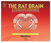
Author: George Paxinos, Charles Watson
Publication date: November 2004 / 209 pages / 5th Edition

The preceding editions made The Rat Brain in Stereotaxic Coordinates the second most cited book in science. Affordable, comprehensive, compact, and convenient, the Fifth Edition is expected to continue the legacy of this major neuroscience publication and be a guide to all students and scientists who study the rat brain. In this edition, for the first time since the first edition, delineations have been drawn entirely anew (on the basis of a new set of sections). Eighteen hitherto unknown nuclei are identified.

Key features:

  • 161 coronal diagrams based on a single brain
  • Delineations drawn entirely new from a new set of sections
  • Diagrams spaced at constant 120 ìm intervals resulting in the high resolution and convenience of use
  • Drawings use blue color lines and black labels to facilitate extraction of information
  • The stereotaxic grid was derived using the same techniques that produced the widely praised stereotaxic grid of the previous editions
  • Over 1000 structures identified, a number for the first time in this edition

Reviews


Review
View All Product Reviews